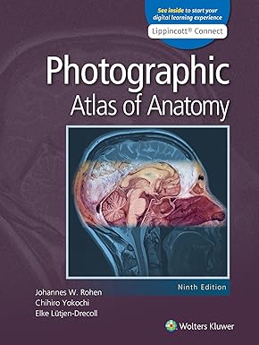
Description
Photographic Atlas of anatomy 9th edition
Product details
- Publisher : LWW
- Publication date : October 10, 2023
- Edition : Ninth
- Language : English
- Print length : 597 pages
- ISBN-10 : 1975229924
- ISBN-13 : 978-1975229924
Photographic Atlas of Anatomy 9th Edition – Real Cadaveric Images for Real Clinical Understanding
When it comes to understanding the human body as it truly looks — not as an artist imagined it — the Photographic Atlas of Anatomy 9th Edition is in a category of its own. Rather than relying on illustrated diagrams, this atlas uses actual cadaveric dissection photographs to show every major structure in the human body with remarkable clarity and authenticity. For students preparing for dissection labs, clinical rotations, or professional exams, it is one of the most honest and practical learning tools available.
While illustrated anatomy books have their place, nothing prepares you for what you will see in the dissection room — or in surgery — quite like photographs of real specimens. That is precisely the gap this atlas fills, and it does so with exceptional quality.
What Does the Photographic Atlas of Anatomy 9th Edition Cover?
This edition provides full photographic coverage of all major regions and structures of the human body, presented in a clean, well-labelled format that is easy to navigate during both study and lab sessions. Each image is carefully dissected and photographed to show structures in their true anatomical relationships.
Coverage includes:
- Head and neck — cranial structures, facial muscles, cervical regions, thyroid, and deep neck dissections
- Thorax — chest wall, lungs, heart, pericardium, mediastinum, and great vessels
- Abdomen — abdominal wall, peritoneal cavity, digestive organs, liver, spleen, kidneys, and retroperitoneum
- Pelvis and perineum — pelvic floor, reproductive organs, bladder, rectum, and perineal structures
- Back and spinal column — vertebral anatomy, spinal cord, meninges, and paraspinal muscles
- Upper limb — shoulder musculature, brachial plexus, elbow and wrist joints, and hand dissections
- Lower limb — hip and knee joints, femoral and popliteal regions, leg compartments, and foot anatomy
- Neuroanatomy — brain and spinal cord sections showing real tissue architecture
The limb and joint photographs are particularly valuable for students working through orthopedic surgery books, offering a level of visual realism that illustrated atlases simply cannot match when it comes to understanding surgical anatomy and tissue planes.
Who Should Use the Photographic Atlas of Anatomy?
This atlas is one of the most practically focused medical textbooks in anatomical education. It serves a wide range of learners and professionals, including:
- MBBS students attending dissection labs — it is an indispensable companion to your core MBBS books, helping you identify structures on cadavers with much greater confidence
- Physiotherapy students who benefit from seeing the true three-dimensional relationships of muscles, nerves, and joints — use it alongside your physiotherapy books to strengthen spatial anatomical understanding
- Orthopedic trainees who need to visualise tissue planes, joint capsule relationships, and neurovascular anatomy as they appear in real dissection and surgical exposure
- Rheumatology students and residents studying the anatomy of synovial joints, periarticular structures, tendon sheaths, and bursae in their actual tissue context
- Surgeons and doctors who want a photographic reference to complement their surgery books and clinical medicine books
- Anyone building a thorough collection of healthcare education resources that reflects the full range of anatomical learning tools
Clinical and Educational Benefits
Photographs Prepare You for the Real Thing
There is a well-known challenge in anatomy education — students who study only from illustrated diagrams are sometimes surprised by what actual tissue looks like during dissection or surgery. The Photographic Atlas of Anatomy closes that gap entirely. By studying from real cadaveric images, you develop visual recognition skills that transfer directly into the lab and the operating room.
This makes it an outstanding complement to illustrated anatomy books and a natural visual reference alongside surgery books that describe anatomical approaches in text.
Supports Deeper Spatial Understanding
Photographs capture depth, texture, colour variation, and tissue relationships in ways that even the best medical illustrations cannot fully convey. For students learning joint anatomy, fascial layers, or neurovascular bundle relationships, seeing these structures photographically accelerates understanding and improves retention significantly.
Valuable Across Multiple Specialties
The atlas carries value well beyond the first year of medical school. Orthopedic trainees revisit limb anatomy before surgical rotations. Rheumatology trainees use it to appreciate periarticular anatomy at a tissue level. Physiotherapy students reference it when studying movement and manual therapy. Its visual honesty makes it useful at every stage of clinical development.
Practical Learning Advantages
Beyond its content, the 9th edition is designed to be genuinely easy to use during study sessions and laboratory work. Key practical advantages include:
- High-resolution cadaveric dissection photographs throughout — updated and refined for the 9th edition
- Clear, consistent labelling on every image for fast, accurate structure identification
- Organised by body region for easy navigation during dissection lab sessions
- Compact and accessible format — easy to carry into the lab or keep open on a desk alongside other texts
- Pairs naturally with any illustrated anatomy atlas or written textbook as a visual reality check
- Covers both gross anatomy and selected neuroanatomy with equal clarity
Key Features of the Photographic Atlas of Anatomy 9th Edition
- ✅ Real cadaveric dissection photographs — not illustrations — throughout every chapter
- ✅ Full regional coverage of the human body from head to foot
- ✅ Updated high-resolution images with clear, accurate anatomical labelling
- ✅ Ideal for dissection lab preparation and clinical anatomy revision
- ✅ Strong visual coverage of joints, fascial planes, and neurovascular anatomy
- ✅ Neuroanatomy section with real brain and spinal cord specimens
- ✅ Natural companion to clinical medicine books, MBBS books, and illustrated atlases
- ✅ Used widely in MBBS, physiotherapy, surgical, and postgraduate anatomy programmes
Frequently Asked Questions (FAQ)
How is a photographic atlas different from an illustrated atlas like Netter?
An illustrated atlas like Netter uses hand-drawn medical illustrations that are anatomically accurate but visually idealised — structures are shown clearly separated and colour-coded for learning purposes. A photographic atlas uses actual cadaveric dissection images, showing tissue exactly as it looks in the real world, including natural colour variation, fat deposits, and structural relationships that illustrations sometimes simplify. Both have real value, and many students use them together for the most complete anatomical understanding.
Is this atlas suitable for MBBS students?
Absolutely. It is particularly useful for first and second year MBBS students attending dissection labs. The photographs help students identify structures on actual cadavers with far greater confidence than diagrams alone allow. It works best alongside your core MBBS books and an illustrated atlas rather than as a standalone anatomy course text.
Is the Photographic Atlas of Anatomy useful for physiotherapy students?
Very much so. Physiotherapy students benefit enormously from seeing the true spatial relationships between muscles, nerves, tendons, and joints — relationships that photography captures far better than diagrams. Used alongside dedicated physiotherapy books, it builds the three-dimensional anatomical thinking that strong clinical reasoning requires.
Can orthopedic and rheumatology trainees use this atlas?
Definitely. The joint dissection photographs — showing knee, shoulder, hip, and wrist anatomy in real tissue — are invaluable for trainees in both specialties. For orthopedic trainees, the tissue plane and surgical anatomy photographs complement orthopedic surgery books in a way that written descriptions and even illustrations cannot replicate. For rheumatology trainees, seeing bursae, tendon sheaths, and periarticular structures photographically reinforces the anatomical basis of inflammatory and degenerative disease.
Does this atlas work well alongside other anatomy resources?
Yes, and that is arguably its best use case. The Photographic Atlas of Anatomy is designed to complement rather than replace illustrated atlases and written anatomy textbooks. Most students find the most effective approach is to read a regional chapter from a written anatomy text, reference an illustrated atlas for diagram clarity, then use this photographic atlas to see how those structures truly appear — closing the loop between theory and reality.
Final Verdict – Why the Photographic Atlas of Anatomy 9th Edition Belongs in Every Serious Medical Library
No other resource prepares you for the reality of anatomy quite like the Photographic Atlas of Anatomy 9th Edition. It does not replace your written texts or illustrated atlases — it completes them. By showing you how the human body truly looks, it transforms theoretical knowledge into genuine visual recognition that you carry with you into every clinical and surgical environment.
Whether you are rounding out your collection of MBBS books, investing in lasting healthcare education resources, or looking for a practical visual companion to your surgery books and clinical medicine books — this atlas earns its place on every serious student’s and clinician’s shelf.
Order the Photographic Atlas of Anatomy 9th Edition today and see human anatomy the way it truly is — no filters, no simplifications, just real anatomy.




Reviews
There are no reviews yet.